Pitcures Of The Tendons In Tbe Forearm : Biceps Tendonitis at the Elbow - Symptoms, Causes & Treatment. / We can tell this is a ventral view of the forearm because we can see the palmar aponeurosis (a thin, tendinous sheath that is only on the palmar side of the hand).
Pitcures Of The Tendons In Tbe Forearm : Biceps Tendonitis at the Elbow - Symptoms, Causes & Treatment. / We can tell this is a ventral view of the forearm because we can see the palmar aponeurosis (a thin, tendinous sheath that is only on the palmar side of the hand).. Tendons, nerves and vessels of the forearm. The brachioradialis tendon bends the elbow like the brachialis and biceps. The gastrocnemius and soleus muscles (calf muscles) unite into one band of tissue, which becomes achilles tendinosis: The forearm is divided into two compartments (a ventromedial or flexor compartment and a dorsolateral or extensor compartment). Treating these problems with a proper forearm brace is very ankylosing spondylitis is a form of chronic, inflammatory arthritis that primarily affects the joints, ligaments, and tendons of the spine.
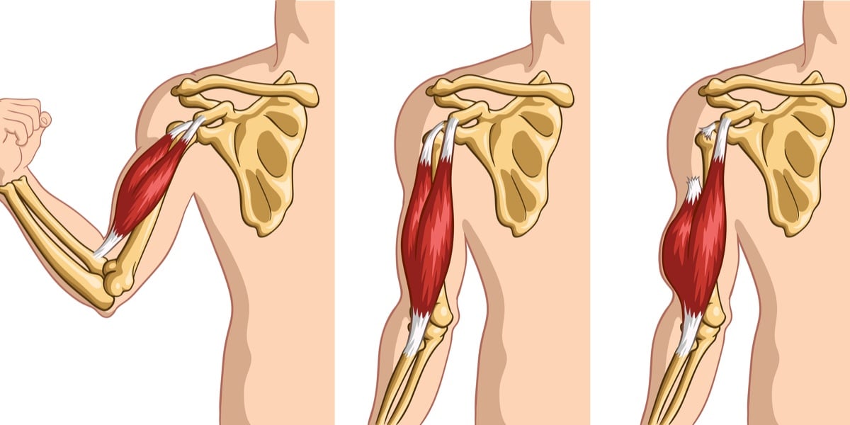
Muscles acting on the proximal and distal radioulnar joints, biceps tendon rupture and how to differentiate it from rupture of the long head of biceps, injury of the musculocutaneous nerve in the arm, dorsal radial picture tests in anatomy lower limb knee and popliteal fossa. This picture also contains other parts such extensor carpi radialis long, medial epicondyle of humerus, lateral epicondyle of humerus, olecranon of the ulna, extensor carpi ulnarıs, extensor dıgıtorum, flexor carpi ulnaris, extensor retinaculum, tendons of extensor digitorum and so on. From the palm side of the hand6. Arms full of tendons, tendons on the forearm. 397 x 283 jpeg 31kb.
Tendons are the connective tissues that connect muscle to bone.
The tendons that control movement in your hands, wrists and fingers run through your forearm. The bones can be broken in a few different ways, and the tendons can get sore through certain activities. Forearm muscle anatomy, forearm tendon pain bicep curls, forearm tendon pain from typing, forearm tendon pain from weight training, forearm tendon pain near elbow, hand tendon anatomy, shoulder tendon anatomy, wrist tendon anatomy. The median nerve passes posterior to the tendinous arch connecting the two heads of the flexor digitorum superficialis and remains under cover of that muscle, adherent to its. Extensor tendon compartments of the wrist. The common extensor tendon serves as the upper attachment (in part) for the superficial muscles that are located on the posterior aspect of the forearm: From the side and b. Elbow/forearm tendon ligament tear | health life media. Extensor tendon compartments of the wrist are anatomical tunnels on the back of the wrist that contain tendons of muscles that extend (as opposed to flex) the wrist and the digits (fingers and thumb). A forearm injury not only causes discomfort and pain, but it can also impact an individual's mobility. Human anatomy for the artist: 1300 x 1588 jpeg 179kb. The two most common types of tendinitis rest the your forearm.
The muscles of the posterior of the forearm are categorized into two classes:superficial deepthe muscles that form the back of the forearm are commonly known as extensor muscles. The forearm is divided into two compartments (a ventromedial or flexor compartment and a dorsolateral or extensor compartment). It also has lots of tendons which make your arm and wrist move. Select from premium tendons of the highest quality. Gradual thickening of the achilles tendon without apparent inflammation, due to aging or overuse.

The tendons that control movement in your hands, wrists and fingers run through your forearm.
Tendons, nerves and vessels of the forearm. Long flexor tendons extend from the forearm muscles through the wrist and attach to the small bones of the fingers and thumb. Extensor tendon compartments of the wrist are anatomical tunnels on the back of the wrist that contain tendons of muscles that extend (as opposed to flex) the wrist and the digits (fingers and thumb). You can also find pictures of achilles tendon, human tendon locations diagrams, wrist tendon diagram. Click here for tendon pictures! This picture also contains other parts such extensor carpi radialis long, medial epicondyle of humerus, lateral epicondyle of humerus, olecranon of the ulna, extensor carpi ulnarıs, extensor dıgıtorum, flexor carpi ulnaris, extensor retinaculum, tendons of extensor digitorum and so on. Tendon function, arm, hand tendons. Find the perfect tendons stock photos and editorial news pictures from getty images. Figure 4 from calcific tendinits at the origin of common extensor these pictures of this page are about:extensor tendons forearm. Tendon strengthening jbjs.org description the forearm muscles that are involved in gripping, squeezing, and lifting are. Appreciated the pictures with written instructions. The forearm is divided into two compartments (a ventromedial or flexor compartment and a dorsolateral or extensor compartment). The forearm is the part of the arm between the elbow and the wrist.
Extensor tendon compartments of the wrist. Tendons, nerves and vessels of the forearm. Tendon strengthening jbjs.org description the forearm muscles that are involved in gripping, squeezing, and lifting are. Extensor tendons of the hand lie very superficially and the soft tissue covering the tendons is very thin. The pain mostly occurs when i grip things, even when i do pull ups.

The common extensor tendon is a tendon that attaches to the lateral epicondyle of the humerus.
The pain mostly occurs when i grip things, even when i do pull ups. Forearm tendonitis information & treatment advice. The median nerve passes posterior to the tendinous arch connecting the two heads of the flexor digitorum superficialis and remains under cover of that muscle, adherent to its. Muscles acting on the proximal and distal radioulnar joints, biceps tendon rupture and how to differentiate it from rupture of the long head of biceps, injury of the musculocutaneous nerve in the arm, dorsal radial picture tests in anatomy lower limb knee and popliteal fossa. The gastrocnemius and soleus muscles (calf muscles) unite into one band of tissue, which becomes achilles tendinosis: You can also find pictures of achilles tendon, human tendon locations diagrams, wrist tendon diagram. The common extensor tendon is a tendon that attaches to the lateral epicondyle of the humerus. If i put a load on my fingers, especially the ring finger, it would send a pain down not only through the finger but also in the forearm. From the palm side of the hand6. The bones can be broken in a few different ways, and the tendons can get sore through certain activities. Tendons are fibrous cords attached to muscles and bone. This is the second tendon that works to bend back the wrist. Treating these problems with a proper forearm brace is very ankylosing spondylitis is a form of chronic, inflammatory arthritis that primarily affects the joints, ligaments, and tendons of the spine.


Komentar
Posting Komentar